We report the anesthesia management of a severe pectus excavatum with cardiac compression displacement under thoracoscopic correction with general anesthesia. Preoperative chest computed tomography showed that the lower end of the sternum was significantly depressed, and the distance between the depressed sternum and the spine was less than 2cm. The heart was obviously compressed and shifted to the left thoracic cavity. During the intraoperative reversal of the orthopedic plate, the blood flow velocity of the tricuspid valve increased to 87.9cm/s and showed a single peak. The transesophageal echocardiography indicated mild tricuspid valve regurgitant flow, and the blood pressure dropped rapidly. After the surgeon was informed of the situation, the orthopedic plate was quickly turned to the convex side up, and the locally depressed anterior chest wall was lifted up. Satisfactory correction of chest wall malformations.
| Published in | American Journal of Pediatrics (Volume 10, Issue 3) |
| DOI | 10.11648/j.ajp.20241003.14 |
| Page(s) | 128-131 |
| Creative Commons |
This is an Open Access article, distributed under the terms of the Creative Commons Attribution 4.0 International License (http://creativecommons.org/licenses/by/4.0/), which permits unrestricted use, distribution and reproduction in any medium or format, provided the original work is properly cited. |
| Copyright |
Copyright © The Author(s), 2024. Published by Science Publishing Group |
Pectus Excavatum, General Anesthesia, Transesophageal Echocardiography, Paravertebral Nerve Block
PE | Pectus Excavatum |
GA | General Anesthesia |
TEE | Transesophageal Echocardiography |
PNB | Paravertebral Nerve Block |
| [1] | Fonkalsrud EW. Open repair of pectus excavatum with minimal cartilage resection [J]. Ann Surg, 2018, 240 (3): 231-235. |
| [2] | PiccoloRL, BonginiU, BasileM, et al. Chest fast MRI: an imaging alternative on pre-operative evaluation of pectus excavatum [J]. J Pediatr Surg, 2012, 47(3): 485-489. |
| [3] | SarwarZU, DeflorioR, O'ConnorSC. Pectus excavatum: current imaging techniques and opportunities for dose reduction [J]. Semin Ultrasound CT MR, 2014, 35(4): 374-381. |
| [4] | LollertA, FunkJ, TietzeN, et al. Morphologic assessment of thoracic deformities for the preoperative evaluation of pectus excavatum by magnetic resonance imaging. [J]. Eur Radiol, 2015, 25(3): 785-791. |
| [5] | AwadSFM, RaimundoBB, BelemLDS, et al. Brugada phenocopy in a patient with pectus excavatum: systematic review of the ECG manifestations associated with pectus excavatum [J]. Ann Noninvasive Electrocardiol, 2013, 18(5): 415-420. |
| [6] | Redlinger RE, Kelly RE, Nuss D, et a1. One hundred patients with recurrent pectus excavatum repaired via the minimally invasive Nuss technique-effective in most regardless of initial operative approach [J]. J Pediatr Surg, 2011•46(6): 1177-1181. |
| [7] | Nuss D, Kelly RE Jr, Croitom DP, et a1. A 10-year review of a minimally invasive technique for the correction of pectus excavatum. J Pediatr Surg. 1998 Apr; 33(4): 545-52. |
| [8] | Nuss D, Kelly RE Jr. Indications and technique of Nuss procedure for pectus excavatum. Thorac Surg Clin, 2018, 20: 583-597. |
| [9] | Matsuo N, Matsumoto K, Taura Y, et al. Initial experience with a 3D printed model for preoperative simulation of the Nuss procedure for pectus excavatum. J Thorac Dis, 2018, 10(2): E120-E124. |
| [10] | Kelly RE, Goretsky MJ, Obermeyer R, et al. Twenty-one years of experience with minimally invasive repair of pectus excavatum by the Nuss procedure in 1215 patients. Ann Surg. 2010; 252(6): 1072-1081. |
| [11] | Mortellaro VE, Iqbal CW, Fike FB, et al. The predictive value of Haller index in patients undergoing pectus bar repair for pectus excavatum. J Surg Res, 2011, 170(1): 104-106. |
| [12] | Gosztyla CE, Petrosyan M, Kane T, et al. Mini thoracic CT adequately determines Haller index and decreases radiation exposure in children with pectus excavatum. J Pediatr Surg, 2022, 57(6): 1076-1078. |
| [13] | Zens TJ, Casar Berazaluce AM, Jenkins TM, et al. The severity of pectus excavatum defect is associated with impaired cardiopulmonary function. Ann Thorac Surg, 2022, 114(3): 1015-1021. |
| [14] | Hebra A, Kelly RE, Ferro MM, et al. Life-threatening complications and mortality of minimally invasive pectus surgery. J Pediatr Surg, 2018, 53(4): 728-732. |
| [15] | Senica SO, Gasparella P, Soldatenkova K, et al. Cardiac perforation during minimally invasive repair of pectus excavatum: A rare complication. J Surg Case Rep, 2022, 2022(11): rjac538. |
| [16] | Dzielicki J, Korlacki W, Janicka I, et al. Difficulties and limitations in minimally invasive repair of pectus excavatum-6 years experiences with Nuss technique [J]. Eur J Cardiothorac Surg, 2006, 30(5): 801-804. |
APA Style
Yang, J., Li, X. (2024). Anesthesia Management of a Child with Severe Pectus Excavatum Complicated with Heart Compression Displacement. American Journal of Pediatrics, 10(3), 128-131. https://doi.org/10.11648/j.ajp.20241003.14
ACS Style
Yang, J.; Li, X. Anesthesia Management of a Child with Severe Pectus Excavatum Complicated with Heart Compression Displacement. Am. J. Pediatr. 2024, 10(3), 128-131. doi: 10.11648/j.ajp.20241003.14
AMA Style
Yang J, Li X. Anesthesia Management of a Child with Severe Pectus Excavatum Complicated with Heart Compression Displacement. Am J Pediatr. 2024;10(3):128-131. doi: 10.11648/j.ajp.20241003.14
@article{10.11648/j.ajp.20241003.14,
  author = {Jiaqi Yang and Xuejie Li},
  title = {Anesthesia Management of a Child with Severe Pectus Excavatum Complicated with Heart Compression Displacement
},
  journal = {American Journal of Pediatrics},
  volume = {10},
  number = {3},
  pages = {128-131},
  doi = {10.11648/j.ajp.20241003.14},
  url = {https://doi.org/10.11648/j.ajp.20241003.14},
  eprint = {https://article.sciencepublishinggroup.com/pdf/10.11648.j.ajp.20241003.14},
  abstract = {We report the anesthesia management of a severe pectus excavatum with cardiac compression displacement under thoracoscopic correction with general anesthesia. Preoperative chest computed tomography showed that the lower end of the sternum was significantly depressed, and the distance between the depressed sternum and the spine was less than 2cm. The heart was obviously compressed and shifted to the left thoracic cavity. During the intraoperative reversal of the orthopedic plate, the blood flow velocity of the tricuspid valve increased to 87.9cm/s and showed a single peak. The transesophageal echocardiography indicated mild tricuspid valve regurgitant flow, and the blood pressure dropped rapidly. After the surgeon was informed of the situation, the orthopedic plate was quickly turned to the convex side up, and the locally depressed anterior chest wall was lifted up. Satisfactory correction of chest wall malformations.
},
 year = {2024}
}
TY  - JOUR
T1  - Anesthesia Management of a Child with Severe Pectus Excavatum Complicated with Heart Compression Displacement

AU  - Jiaqi Yang
AU  - Xuejie Li
Y1  - 2024/08/06
PY  - 2024
N1  - https://doi.org/10.11648/j.ajp.20241003.14
DO  - 10.11648/j.ajp.20241003.14
T2  - American Journal of Pediatrics
JF  - American Journal of Pediatrics
JO  - American Journal of Pediatrics
SP  - 128
EP  - 131
PB  - Science Publishing Group
SN  - 2472-0909
UR  - https://doi.org/10.11648/j.ajp.20241003.14
AB  - We report the anesthesia management of a severe pectus excavatum with cardiac compression displacement under thoracoscopic correction with general anesthesia. Preoperative chest computed tomography showed that the lower end of the sternum was significantly depressed, and the distance between the depressed sternum and the spine was less than 2cm. The heart was obviously compressed and shifted to the left thoracic cavity. During the intraoperative reversal of the orthopedic plate, the blood flow velocity of the tricuspid valve increased to 87.9cm/s and showed a single peak. The transesophageal echocardiography indicated mild tricuspid valve regurgitant flow, and the blood pressure dropped rapidly. After the surgeon was informed of the situation, the orthopedic plate was quickly turned to the convex side up, and the locally depressed anterior chest wall was lifted up. Satisfactory correction of chest wall malformations.

VL  - 10
IS  - 3
ER  -
Department of Anesthesiology, West China Hospital, Sichuan University and The Research Units of West China (2018RU012), Chinese Academy of Medical Sciences, Chengdu, China
Department of Anesthesiology, West China Hospital, Sichuan University and The Research Units of West China (2018RU012), Chinese Academy of Medical Sciences, Chengdu, China
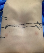
Figure 1. Marked depression in the anterior lower chest.
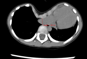
Figure 2. Preoperative chest CT showed significant depression at the lower end of the sternum. The minimum distance between the depressed sternum and the spine was less than 2cm, and the heart was obviously compressed and shifted to the left thoracic cavity.
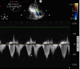
Figure 3. Transesophageal echocardiography examination before operation showed a bimodal flow velocity of 66.7cm/s across the tricuspid valve.
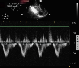
Figure 4. In the process of reversing the orthopedic plate, the flow velocity of the tricuspid valve increased to 87.9cm/s and showed a unimodal peak.
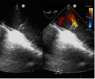
Figure 5. Transesophageal echocardiography indicates mild tricuspid regurgitation during orthosis reversal.
Information